The abdomen describes a portion of the trunk connecting the thorax and pelvis. An abdominal wall formed of skin, fascia, and muscle encases the abdominal cavity and viscera.
Anterolateral Abdominal Wall Muscles
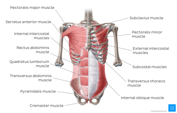
Anterior chest wall strength and movement receive contributions laterally by three layers of large flat paired muscles: the external oblique, internal oblique, and transversus abdominis. Anteromedially these layers fuse to form a rectus sheath that encloses the rectus abdominis and pyramidalis muscles. In the midline, the combined aponeuroses of these muscles fuse to form the linea alba.
External Oblique
The external oblique is the most superficial of the anterolateral abdominal wall muscles. Its fibers arise from the fifth through twelfth ribs and run inferomedially. As it approaches the midclavicular line, its fibers form an aponeurotic sheath, which travels superficially across the rectus abdominis to the linea alba in the midline. Together with the internal oblique, contraction causes rotation and lateral flexion of the vertebral column. Its inferior border forms the inguinal ligament, which runs between the ASIS and pubic tubercle.
Internal Oblique
This muscle lies immediately deep to external oblique. Together with the external oblique, contraction causes rotation and lateral flexion of the vertebral column. It originates from the lumbar fascia, iliac crest, and lateral inguinal ligament. Its fibers run superomedially, orthogonal to the external oblique before also becoming aponeurotic. Medially, its contributions to the rectus sheath differ between its upper fibers and lower fibers. Upper fibers divide to enclose the rectus sheath anteriorly and posteriorly. Inferiorly, all fibers travel anterior to the rectus abdominal muscle. This portion of the rectus sheath will be deficient posteriorly, with no aponeurotic layer between the rectus abdominis and the transversalis fascia. The inferior end of the posterior rectus sheath is called the “arcuate line.” Irrespective of their relationship to rectus abdominis, all layers continue medially to join the linea alba in the midline.
Transversus Abdominis
This muscle is the deepest of the anterolateral muscles. It arises from the fifth through tenth costal cartilages, lumbar fascia, iliac crest, and lateral inguinal ligament. Its fibers run transversely before becoming aponeurotic and running into the rectus sheath. Its upper portion travels posteriorly to the rectus abdominal muscles, contributing to the posterior rectus sheath. Below the arcuate line, its aponeurosis runs anteriorly to the muscle, contributing to the anterior sheath. The contraction of the transversus abdominis causes compression of abdominal contents. The inferior edge of the internal oblique and transversus abdominis form the conjoint tendon.
Rectus Abdominis
This muscle is long and narrow, running in the rectus sheath vertically and parallel to the linea alba. It arises from the pubic symphysis and crest and runs superiorly to attach to the fifth through seventh costal cartilages. It is a powerful flexor of the vertebral column. Each muscle belly is divided by three tendinous intersections into four discrete muscle segments. The tendinous intersections have a tether to the overlying anterior rectus sheath. In an athlete, these may be visible and described as a ‘six-pack’.
Pyramidalis
This muscle is a minor triangular muscle that sits anteriorly to the rectus abdominis in the inferior rectus sheath. Its origin is from the body of the pubis and tenses the linea alba when contracting. It is present bilaterally in 80% of people.
Posterior Abdominal Wall Muscles
Quadratus Lumborum
This muscle originates from the iliolumbar ligament and iliac crest and runs superomedially to insert into the twelfth rib and L1-4 transverse processes. Contraction can cause lateral flexion and extension of the vertebral column, and depression of the rib cage.
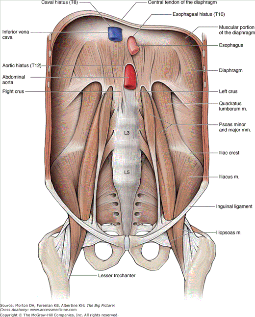
Psoas Major
This muscle originates from the transverse processes of T12 and L1-4 vertebrae and lateral surfaces of the intervening intervertebral discs. It runs inferiorly, joining the iliacus to insert into the lesser trochanter of the femur. Contraction causes flexion of the thigh.
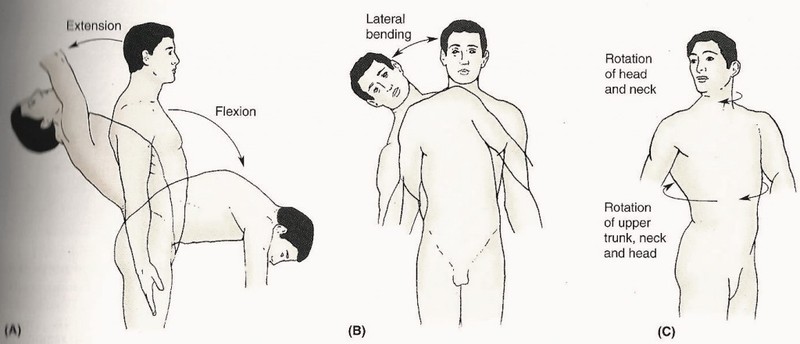
The Left figure shows trunk in flexion
Weakness leads to
- Improper posture
- Specific areas stiffer
- Specific areas overstretched
Strengthen by
- Lumbar flexion
- Building muscle strength
- Relaxing the stiffness
- Destraining the overstretched